Cell culture work supports many of today's core research workflows, from protein expression and imaging assays to drug screening and basic cell biology. A healthy culture gives clean data and confident interpretation, so everyday technique matters as much as the experiment design. Cell culture contamination is one of the most common reasons for unexpected cell behavior, poor growth, or inconsistent results, and it can spread quickly when cultures share reagents, hoods, incubators, or water baths. The most reliable approach is a prevention mindset: build strong routines, use consistent aseptic practices, and verify culture quality on a schedule.
This guide explains practical steps to prevent cell culture contamination with clear, bench-ready advice. You will learn aseptic cell culture techniques, how to spot early warning signs, and how to reduce risk from common sources like hands, aerosols, shared bottles, incubators, and pipettes. We will also cover the two high-impact problems labs track closely: bacterial contamination in cell culture and mycoplasma contamination.
What is cell culture contamination?
Cell culture contamination happens when unwanted organisms or cross-contaminating cells enter a culture system. The most common contaminants include:
- Bacteria (often fast-growing and easy to notice)
- Fungi and yeast (visible filaments or cloudy media)
- Mycoplasma (often hard to see and easy to overlook)
- Cross-contamination with other cell lines (a major data integrity risk)
Contamination changes growth rate, metabolism, gene expression, and experimental readouts. A culture can appear "fine" and still harbor mycoplasma, which is why routine checks are essential for strong cell culture sterility.
Why contamination happens: common lab sources
Most contamination sources connect to a few patterns. When you understand the patterns, prevention becomes predictable.
1) Hands, gloves, and contact transfer
-
Gloves touch door handles, notebooks, pens, keyboards, and then touch sterile tubes.
2) Aerosols and splashes
-
Quick pipetting can generate aerosols.
3) Shared reagents and multi-user bottles
- Shared media, PBS, trypsin, and supplements can become "community sources."
- A single contaminated bottle can affect many cultures.
4) Incubators, water pans, and water baths
- Humidity pans can grow microbes when not cleaned.
- Water baths can transfer organisms to the surfaces of tubes.
Aseptic cell culture techniques that reduce contamination
Strong aseptic cell culture techniques rely on consistent actions that support sterile airflow, clean surfaces, and controlled handling.
Work surface and hood setup
- Clean the biosafety cabinet (BSC) work surface before and after use.
- Arrange items in the hood to keep a clean-to-dirty flow.
- Keep the center workspace clear to foster an open culture.
- Place waste to one side and avoid reaching over open vessels.
Glove and hand habits
- Wear fresh gloves for cell culture work.
- Spray or wipe gloves with 70% ethanol, then allow a brief contact time.
- Change gloves after touching non-sterile surfaces.
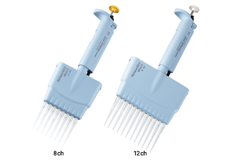
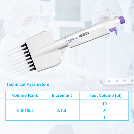
Pipetting technique
- Use sterile, filtered tips for cell culture.
- Pipette smoothly to reduce aerosols.
- Avoid touching pipette tips to non-sterile surfaces.
Bottle and tube handling
- Open bottles and tubes inside the hood, away from the front edge.
- Hold caps facing down or sideways, never facing up.
- Close containers quickly and avoid long "open" time.
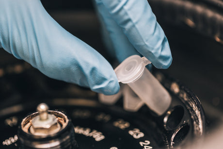
Smart batching
- Prepare aliquots of supplements and reagents to limit repeated bottle entries.
- Use single-use aliquots for high-risk items like FBS, antibiotics, and growth factors.
Preventing cell culture contamination: a practical checklist
Use this prevention checklist as a routine you can teach to new team members.
Daily practices
- Confirm the hood is running and the workspace is clear.
- Wipe surfaces and gloves before handling cultures.
- Label everything before opening containers.
- Keep only essential items in the hood.
- Keep the incubator door open for the shortest practical time.
Weekly practices
- Clean hood surfaces more thoroughly, including the sash and side walls.
- Wipe incubator shelves and door surfaces.
- Check the humidity pan condition and refill with sterile water, as per your SOP.
Monthly practices
- Deep-clean incubators according to manufacturer guidelines.
- Review culture logs for pattern signals (repeat issues on specific days or users).
- Confirm mycoplasma testing schedule is being followed.
These steps support strong cell culture sterility and make prevention a predictable system.
Early signs of contamination: what to watch for
A culture often shows subtle changes before it becomes obviously contaminated.
Visual signs in media
- Unexpected turbidity or a "sparkly" look under the microscope
- Floating particles that move quickly (common with bacteria)
- Filaments, clumps, or web-like structures (common with fungi)
- Sudden color shift in the pH indicator without a clear reason
Cell behavior signs
- Faster or slower growth than usual
- Cells detach more easily than expected
- Increased debris or granularity
- Changes in morphology that do not match the experiment condition
Bench note: Mycoplasma contamination often shows as slower growth, stressed morphology, or inconsistent transfection results. Routine testing makes detection dependable.
Bacterial contamination in cell culture: prevention and response
Bacterial contamination in cell culture often appears quickly. It can cause cloudy media and fast-moving particles under the microscope.
Common sources
- Non-sterile technique during pipetting or bottle opening
- Contaminated water bath surfaces or incubator humidity pans
- Shared reagent bottles
Prevention steps that work
- Use filtered tips and avoid aerosols.
- Store media and reagents properly and avoid prolonged exposure to room temperature.
- Aliquot shared reagents and label with open date.
- Keep incubator and hood cleaning consistent.
Practical response approach
When bacteria appear, fast containment protects other cultures.
- Seal contaminated flasks/plates and remove them carefully.
- Clean the hood workspace after handling.
- Check shared reagents used with that culture.
- Review incubator conditions and clean surfaces.
This approach supports lab-wide consistency and keeps contamination from spreading.
Mycoplasma contamination: why labs take it seriously
Mycoplasma contamination is common in cell culture environments because mycoplasma are small and may not cause obvious turbidity. They can alter gene expression, metabolism, and cell signaling, which changes experimental outcomes.
Typical signs
- Gradual decline in growth
- Lower transfection efficiency
- Increased cell stress and debris
- Inconsistent assay signals across replicates
Best prevention strategy
- Test routinely, even when cultures look healthy.
- Quarantine new cell lines until they pass screening.
- Keep separate media and reagents for high-value lines.
- Use dedicated pipettes or a dedicated hood zone when possible.
Testing basics
Many labs use:
- PCR-based kits for Mycoplasma DNA
- Enzymatic assays
- DNA staining-based methods
Choose a method that fits your throughput and SOP. Consistent schedules build reliable cell culture sterility.
Table: contamination types, signs, and prevention actions
|
Contamination type |
Common signs |
Prevention that helps most |
|
Bacteria |
Cloudy media, fast-moving dots |
Filtered tips, quick containment, clean incubator/humidity pan |
|
Fungi/yeast |
Filaments, clumps, floating “flakes” |
Strong hood discipline, minimize open time, clean shared areas |
|
Mycoplasma |
Subtle stress, slow growth, assay drift |
Routine testing, quarantine new lines, dedicated reagents |
|
Cross-contamination (cell lines) |
Unexpected marker expression, odd morphology |
Authentication, careful labeling, one line open at a time |
Avoiding cell culture contamination through workflow design
Good technique becomes even stronger when the workflow layout supports it.
Separate zones
- Use a clean zone for unopened sterile supplies.
- Use a working zone in the hood for open cultures.
- Use a separate area for waste handling and labeling.
One culture open at a time.
This single habit reduces cross-contamination risk and improves focus.
Label first, then open.
Labeling early reduces mix-ups and helps culture tracking.
Reduce shared bottle entries.
Aliquots reduce the chance that a single mistake affects many lines.
Build a culture log.
A simple log supports pattern detection:
- Cell line name, passage number, split ratio
- Media lot and supplement lot
- Incubator used
- Mycoplasma test date and result
Antibiotics in cell culture: helpful tool with clear limits
Antibiotics can support routine workflows, especially for short periods or when handling new lines, and many labs use them as a practical layer of protection.
At the same time, antibiotics work best as a supplement to aseptic technique rather than a replacement.
Practical habits that keep antibiotic use effective:
- Use antibiotics consistently only when your SOP supports it.
- Keep aseptic technique strong even when antibiotics are present.
- Schedule antibiotic-free windows for sensitive assays if needed.
- Prioritize Mycoplasma testing because standard antibiotics often do not address it.
This balanced approach strengthens data confidence and supports long-term culture quality.
Incubator and hood care that supports cell culture sterility
Biosafety cabinet (BSC) habits
- Keep airflow clear by avoiding clutter.
- Clean surfaces before and after work.
- Keep items off the rear grill and do not block vents.
CO₂ incubator habits
- Wipe door surfaces regularly.
- Clean shelves on a schedule.
- Maintain humidity pan hygiene in accordance with your SOP.
- Monitor temperature and CO₂ stability.
Water bath habits
- Keep lids closed when possible.
- Change water on a schedule and keep surfaces clean.
- Avoid placing open tubes directly near bath splashes.
These routines reduce contamination sources and strengthen the prevention of cell culture contamination across the lab.
Troubleshooting contamination patterns
When contamination repeats, a structured review helps you find the source quickly.
Questions that often reveal the cause
- Does contamination happen in one incubator more than others?
- Does it happen after using a specific shared reagent bottle?
- Does it appear after a specific procedure, like thawing, passaging, or transfection?
- Does it cluster on certain days or users?
Practical investigation steps
- Quarantine cultures and replace shared reagents in a controlled way.
- Clean the incubator and hood, then reintroduce cultures with fresh aliquots.
- Review technique consistency and glove habits.
- Verify mycoplasma testing and cell line identity.
A calm, repeatable process makes avoiding cell culture contamination easier over time.
FAQs
What is the most common cause of cell culture contamination?
The most common cause is contact transfer from hands, gloves, and frequently touched surfaces. Small lapses—like touching a notebook or incubator handle and then handling sterile caps—can introduce organisms that spread through shared reagents and incubators.
How can I tell if my cell culture has bacterial contamination?
Bacterial contamination often shows as cloudy media and many tiny, fast-moving particles under the microscope. Cultures may also show rapid pH changes and stressed cell morphology.
Why is mycoplasma contamination hard to detect?
Mycoplasma are very small and often do not make media cloudy. They can change cell growth and assay performance without obvious visual signs, so routine testing is the most dependable way to detect them.
How often should I test for mycoplasma contamination?
Many labs test routinely on a set schedule and also screen all new or thawed lines before mixing them into shared incubators. A consistent cadence that matches your lab's throughput maintains cell culture sterility.
Do antibiotics prevent cell culture contamination?
Antibiotics can reduce the impact of some bacterial contaminants, but they do not replace aseptic cell culture techniques. They can also mask low-level contamination and they do not reliably address mycoplasma, so testing and prevention habits remain essential.
Conclusion
Strong cell culture results come from consistent technique and a prevention-first mindset. Aseptic cell culture techniques keep exposure low, and smart workflow design reduces the risk that a single event affects multiple cultures. Routine cleaning, careful reagent handling, and a clear testing schedule support dependable cell culture sterility. With these habits in place, preventing cell culture contamination becomes standard practice, and mycoplasma contamination is easier to detect and control before it affects your data.