Cell culture has gone through a quiet evolution. For a long time, almost every experiment was done in flat, two-dimensional (2D) monolayers. Now, three-dimensional (3D) cell culture is showing that cells behave very differently when they’re allowed to grow in a more realistic, tissue-like environment. That’s why people search for “2D vs 3D cell culture,” “2D vs 3D cell culture models,” “2D vs 3D cell culture for drug screening,” and even specific use cases like “2D vs 3D cell culture cardiomyocytes.”
This article explains, what each approach actually is, where each one works best, how 3D methods are set up in the lab.
2D vs 3D Cell Culture: What’s the Core Difference?
When people say “cell culture 2D vs 3D,” they’re really asking: how different is a flat monolayer from a mini 3D micro-tissue?
- 2D cell culture (also called 2D monolayer cell culture or 2D adherent cell culture) grows cells on a flat, treated plastic surface. Cells spread, attach, and form a single layer.
- 3D cell culture lets cells grow in all directions — as spheroids, organoids, or within a scaffold. Cells interact with each other and with a matrix, and gradients of oxygen and nutrients can form.
In the body, cells do not live as flat sheets on plastic. So 3D culture is often better for modeling physiological events in 2D vs 3D cell culture, especially for cancer, drug penetration, and stem cell differentiation. But 2D is still far easier, cheaper, and more reproducible. That’s why you need to understand both.
What Is 2D Cell Culture?
2D cell culture is the traditional, textbook method. You seed cells into a flask, dish, or even a small format like a 2D cell culture 3-well plate. Cells attach to the bottom, spread out, and grow as a uniform monolayer. Most introductory courses, SOPs, and older publications are based on this method.

2D Cell Culture Advantages and Disadvantages
Advantages
- Very easy to learn and standardize.
- Inexpensive: basic plasticware and standard cell culture media are enough.
- Ideal for high-throughput experiments.
- Cells are easy to image with standard microscopes.
- Perfect for routine 2D cancer cell culture, transfections, and viability assays.
Disadvantages
- Cell morphology is altered — cells flatten more than they would in vivo.
- Limited cell–cell and cell–matrix interactions.
- Drug responses can be less predictive than in 3D models.
- Some physiological processes cannot be modeled accurately (invasion, complex differentiation).
- You quickly run into the classic “2D cell culture limitation” when you try to replicate tissue-like behavior.
Variants and Special Cases
- 2D cell culture protocol: usually seeding → attachment → media change → passage. Very standardized.
- 2D cell culture history: 2D became dominant because plastics, incubators, and early cell lines were all designed around it.
- 2D cell culture plants: plant cells can be grown in 2D, but many plant systems shift to callus or suspension culture.
- 2D adherent cell culture vs suspension: most mammalian 2D systems are adherent, but some lines can be adapted to suspension if needed.
So, 2D remains the “default” because it’s predictable.
What Is 3D Cell Culture?
3D cell culture is any in vitro method that lets cells grow in three dimensions so they form structures closer to real tissues. Instead of a flat monolayer, you get spheroids, clusters, or cells embedded in a matrix. This is why researchers write “2D vs 3D cell culture review” papers — moving from 2D to 3D changes how cells behave, proliferate, and respond to drugs.

3D Cell Culture Advantages and Disadvantages
Advantages
- More physiologically relevant — closer to in vivo.
- Better for tumor, immuno-oncology, and organoid work.
- Drug diffusion and penetration can be studied realistically.
- Good for modeling physiological events in 2D vs 3D cell culture such as hypoxia and ECM interactions.
- Pairs well with 3D cell culture imaging to study structure.
Disadvantages
- Protocols are longer and more technical.
- Not every lab has experience with 3D cell culture methods.
- Spheroids can vary in size, making quantification harder.
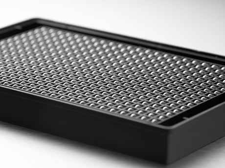
- Requires specialized consumables like 3D cell culture plates or matrices.
- Sometimes you need instruments for automated 3D cell culture screening if the workflow is large.
Common 3D Cell Culture Techniques
-
3D cell culture spheroids
Cells are cultured in low-attachment plates or U-bottom wells so they self-aggregate. This is a widely used, relatively simple 3D model. -
Scaffold-based 3D cell culture methods
Cells are grown within or on a biomaterial that mimics extracellular matrix. -
Hanging drop method
Often listed as “e hanging drop method 3D cell culture model protocols.” Cells are placed in small drops; gravity pulls them together to form spheroids. -
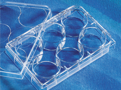
3D cell culture plates
Vendors (for example, Corning 3D cell culture products) make plates whose geometry or coating promotes even 3D growth. -
Automated 3D cell culture
For high-throughput labs, automated systems seed, feed, and read 3D models so they can be used for screening.
2D vs 3D Cell Culture Models: Where Each One Fits
2D vs 3D Cell Culture for Drug Screening
- 2D is still the workhorse for large-scale drug screening because you can plate hundreds or thousands of wells quickly and get clean, comparable data.
- 3D is more informative when you want to know: will this compound actually reach inner cells, will the tumor-like structure resist it, and will it behave more like in vivo? That’s why many groups run: primary screen in 2D → confirm and profile in 3D.
So the answer is not “2D or 3D,” it’s “2D first, 3D for validation.”
2D vs 3D Cell Culture Cardiomyocytes
Cardiomyocytes in 2D can attach and even beat, but the alignment, electrical coupling, and 3D architecture are limited. In 3D, these cells can form more tissue-like constructs and respond in a way that is closer to heart tissue. That’s why the query “2d vs 3d cell culture cardiomyocytes” exists — people see a real functional difference.
2D vs 3D Cell Culture Nanoparticles
Nanoparticle uptake and distribution can look great in 2D but drop in 3D because of diffusion barriers and layered cell structures. If the goal is to study delivery or penetration, 3D is the more realistic model.
Modeling Physiological Events in 2D vs 3D Cell Culture
Processes that depend on gradients, ECM, or 3D contacts — such as invasion, metastasis modeling, or oxygen-limited growth — are simply better in 3D. Processes that are more about pathways, reporter genes, or basic viability can stay in 2D.
Lab Setup for 3D Cell Culture
A lot of people search “3D cell culture techniques” and “3D cell culture methods” but then realize they don’t know what to buy. Here’s a practical view.

What You Probably Already Have
- Biosafety cabinet
- CO₂ incubator
- Standard pipettes
- Inverted microscope
You can do basic 3D work with just these.
What You Add for 3D
-
3D cell culture plates
Ultra-low attachment or U-bottom plates help form uniform spheroids. These are important if you want consistent size for imaging or screening. -
Matrices or hydrogels
For scaffold-based 3D, you’ll need ECM-like materials. -
Written 3D cell culture protocol
3D needs more precise seeding densities and media change schedules than 2D. -
Imaging plan
3D cell culture imaging can be done with standard microscopes at first, but confocal or advanced imaging helps if your model is thick.
Automated 3D Cell Culture and Screening
This is a growing part of the 3D cell culture market. High-throughput 3D is now possible, especially in pharma and CRO environments. This is also the kind of thing that appears at events like “3d cell culture conference 2025,” where vendors show systems that integrate seeding, incubation, and readouts.
2D vs 3D Cell Culture Review: When to Use Which

- Use 2D cell culture when you want speed, low cost, established protocols, student training, or high-throughput drug screening.
- Use 3D cell culture when you want physiological relevance, tumor-like behavior, diffusion or penetration studies, cardiomyocyte or stem-cell differentiation models, or when your 2D data does not match animal/clinical data.
You can also mix the two in a single project: screen in 2D, confirm in 3D, publish with both to show robustness.
Real Queries
- 2d vs 3d cell culture ppt: this topic translates very well into slides — definition → comparison table → applications → lab setup.
- 3d cell culture conference 2025: shows that the field is active and vendors are pushing toward automation and organoid-style models.
- corning 3d cell culture: a concrete example of commercially available 3D tools — good to mention in methods sections.
- 3d cell culture market: signals that demand for 3D consumables and instruments is rising, so labs should start learning these techniques now.
Commonly Asked Questions
What is the main difference between 2D and 3D cell culture?
2D cell culture grows cells as a flat monolayer on plastic; 3D cell culture allows cells to grow in all directions, forming spheroids or tissue-like structures that behave more like cells in the body.
Is 3D cell culture always better?
No. 3D is more realistic, but it is also more work. For fast, repeatable assays, 2D is still the best choice.
What are 3D cell culture plates?
These are specially designed plates with low-attachment or U-bottom wells that promote formation of uniform 3D spheroids.
Can 3D cell culture be automated?
Yes. Automated 3D cell culture and automated 3D cell culture screening systems exist and are being adopted where high throughput is needed.
Why do drug screening labs move to 3D?
Because drug penetration and resistance in 3D can be closer to in vivo tumors than in 2D monolayers, giving more predictive data.
What is the hanging drop method in 3D cell culture?
It’s a simple protocol where cells are placed in small drops and allowed to aggregate by gravity, producing spheroids without special plates.
Are 2D cell culture protocols still relevant?
Absolutely. 2D remains the foundation of cell culture training and many validated assays are still based on 2D.
Can I use 2D and 3D in the same study?
Yes. Many good studies show primary data in 2D and confirm key findings in 3D.
What is “2d vs 3d cell culture review” that I see in search?
It’s usually an academic-style paper that compares both systems across morphology, gene expression, drug response, and practical considerations.
Why are there searches for “3d cell culture conference 2025” and “3d cell culture market”?
Because 3D is becoming commercialized — vendors, researchers, and pharma companies are investing in better 3D platforms and sharing updates at conferences.